Anatomy Of Chest Wall / Axial Muscles Of The Abdominal Wall And Thorax Anatomy And Physiology - This chapter is an abbreviated review of thoracic anatomy as seen on chest.. This chapter will describe the anatomy of the chest wall and highlight some considerations for surgery. The bony skeletal part of the thoracic wall is the rib cage, and the rest is made up of muscle, skin, and fasciae. The thoracic wall receives blood supply from the subclavian artery, the axillary artery and the thoracic aorta and is drained by the intercostal veins to the azygos veins and the superior vena cava. Course transversely along the superior portion of the chest wall from the manubrium to the acromion of the scapula and, therefore, are easily palpable. The chest wall mass was biopsied and confirmed to be a chondrosarcoma.
Jugular notch, sternoclavicular joint, superior border of. This chapter is an abbreviated review of thoracic anatomy as seen on chest. The chest wall is comprised of skin, fat, muscles, and the thoracic skeleton. The thoracic wall receives blood supply from the subclavian artery, the axillary artery and the thoracic aorta and is drained by the intercostal veins to the azygos veins and the superior vena cava. Actually we see the interface between the air in the lungs and the soft tissue structures in the abdomen.
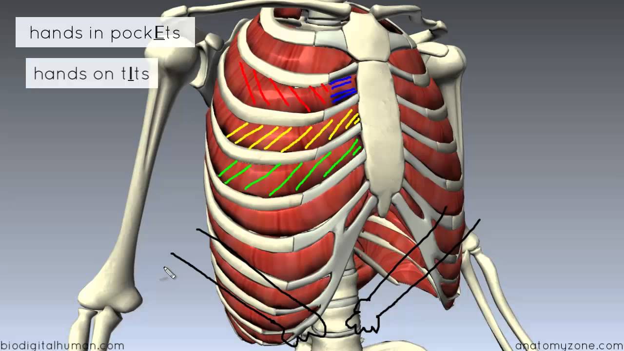
Muscles Of The Thoracic Wall 3d Anatomy Tutorial Youtube from i.ytimg.com Anterior thoracic wall the anterior thoracic wall extends craniocaudally from the level of the clavicle and jugular notch to the level of the xiphisternal joint. Bony thorax in a cadaver: The skeleton of the thoracic wall is formed by the twelve thoracic vertebra posteriorly, the sternum anteriorly and, on each side, by the twelve ribs and the respective costal cartilage. Use the mouse scroll wheel to move the images up and down alternatively use the tiny arrows (>>) on both side of the image to move the images.>>) on both side of the image to move the images. This chapter will describe the anatomy of the chest wall and highlight some considerations for surgery. Thoracic wall dissection anatomy description: The dominant muscle in the upper chest is the pectoralis major. The chest wall functions as a protective cage around the vital organs of the body, and significant disruption of its structure can have dire respiratory and circulatory consequences.
The chest wall, like other regional anatomy, is a remarkable fusion of form and function.
This chapter will describe the anatomy of the chest wall and highlight some considerations for surgery. Bony thorax in a cadaver: The dominant muscle in the upper chest is the pectoralis major. Anatomy of the thoracic wall. The spaces between the ribs are filled by the intercostal musculature, which consists of three layers. Actually we see the interface between the air in the lungs and the soft tissue structures in the abdomen. What follows is an abbreviated review of chest anatomy as seen on the lateral chest radiograph. It provides protection to vital organs (eg, heart and major vessels, lungs, liver) and provides stability for movement. Course transversely along the superior portion of the chest wall from the manubrium to the acromion of the scapula and, therefore, are easily palpable. Chest wall anatomy the chest is considered to be the area between the neck and the abdomen and contains many major organs as well as muscle groups. The left diaphragm can only be seen to a point where it borders the heart (blue arrow). Some of the chest wall muscles can be used as helpful anatomical landmarks. Retaining ligaments of the lateral chest wall suspend the lateral portion of the breast parenchyma and are often divided during a mastectomy.
It is made up of the manubrium superiorly, the body and the xiphisternum (figure 1). Anatomy of the thoracic wall. The mammary gland is located within the superficial fascia of the anterior thoracic wall. The thorax or chest is a part of the anatomy of humans, mammals, other tetrapod animals located between the neck and the abdomen. Learn about chest wall anatomy.
Chest Wall Anatomy Springerlink from media.springernature.com Overview of the components of the thoracic wall. The chest wall is formed from the sternum anteriorly, 12 pairs of ribs, costal cartilages and intercostal muscles laterally, and the thoracic vertebrae posteriorly. However, the clavicle, overly rib 1, which makes rib 1 impalpable. Actually we see the interface between the air in the lungs and the soft tissue structures in the abdomen. Computed tomography (ct) of the chest can detect pathology that may not show up on a conventional chest radiograph(1). 2 skin of the anterior chest wall syllabus p. Bony thorax in a cadaver: The chest wall, like other regional anatomy, is a remarkable fusion of form and function.
The mammary gland is located within the superficial fascia of the anterior thoracic wall.
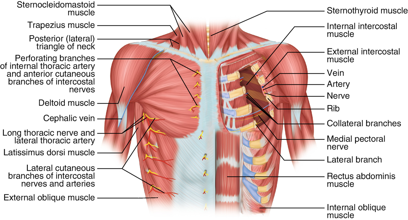
As the heart pumps inside the center of the chest,. The palpable midline sternum is variable in size and shape; The chest wall is formed from the sternum anteriorly, 12 pairs of ribs, costal cartilages and intercostal muscles laterally, and the thoracic vertebrae posteriorly. The superior thoracic aperture found superiorly and the inferior thoracic aperture located inferiorly. The chest wall is comprised of skin, fat, muscles, and the thoracic skeleton. Overview of the components of the thoracic wall. The chest wall has 10 layers, namely (from superficial to deep) skin (epidermis and dermis), superficial fascia, deep fascia and the invested extrinsic muscles (from the upper limbs), intrinsic muscles associated with the ribs (three layers of intercostal muscles. The mammary gland is located within the superficial fascia of the anterior thoracic wall. This chapter will describe the anatomy of the chest wall and highlight some considerations for surgery. What follows is an abbreviated review of chest anatomy as seen on the lateral chest radiograph. Anterior thoracic wall the anterior thoracic wall extends craniocaudally from the level of the clavicle and jugular notch to the level of the xiphisternal joint. 2 skin of the anterior chest wall syllabus p. Surface anatomy of anterior chest wall.
Use the mouse scroll wheel to move the images up and down alternatively use the tiny arrows (>>) on both side of the image to move the images.>>) on both side of the image to move the images. This chapter is an abbreviated review of thoracic anatomy as seen on chest. The chest wall functions as a protective cage around the vital organs of the body, and significant disruption of its structure can have dire respiratory and circulatory consequences. Course transversely along the superior portion of the chest wall from the manubrium to the acromion of the scapula and, therefore, are easily palpable. The chest wall is supplied by the posterior intercostal arteries arising from the aorta, the internal thoracic and the highest intercostals given off the subclavian artery, and the branches of the axillary artery (fig.
Thoracic Wall And Breast Illustrations from www.imaios.com The thorax has two major openings: Principal functions are the protection of internal viscera and an expandable cylinder facilitating variable gas flow into the lungs. It provides protection to vital organs (eg, heart and major vessels, lungs, liver) and provides stability for movement. The bony skeletal part of the thoracic wall is the rib cage, and the rest is made up of muscle, skin, and fasciae. The mammary gland is located within the superficial fascia of the anterior thoracic wall. The right diaphragm should be visible all the way to the anterior chest wall (red arrow). Anatomy of chest wall and mechanics of breathing able to describe the anatomy of the pleural cavity the pleural cavity is as if the lungs have been pushed into. Surface anatomy of anterior chest wall.
The palpable midline sternum is variable in size and shape;
Located between the medial ends of the clavicles. Anatomy of chest wall and mechanics of breathing able to describe the anatomy of the pleural cavity the pleural cavity is as if the lungs have been pushed into. The left diaphragm can only be seen to a point where it borders the heart (blue arrow). The chest wall is comprised of skin, fat, muscles, and the thoracic skeleton. Course transversely along the superior portion of the chest wall from the manubrium to the acromion of the scapula and, therefore, are easily palpable. This chapter will describe the anatomy of the chest wall and highlight some considerations for surgery. The dominant muscle in the upper chest is the pectoralis major. 30 lines of the thoracic wall syllabus p. The chest wall, like other regional anatomy, is a remarkable fusion of form and function. The bony skeletal part of the thoracic wall is the rib cage, and the rest is made up of muscle, skin, and fasciae. The chest or thorax is the region between the neck and diaphragm that encloses organs, such as the heart, lungs, esophagus, trachea, and thoracic diaphragm. The right diaphragm should be visible all the way to the anterior chest wall (red arrow). Anatomy of the thoracic wall.
The skeleton of the thoracic wall is formed by the twelve thoracic vertebra posteriorly, the sternum anteriorly and, on each side, by the twelve ribs and the respective costal cartilage anatomy of chest. 4 innervation of the breast blood supply of the breast syllabus p.